The SMR Map
Self Myofacial Release (SMR): A self massage technique to relieve tightness
How to Use THe SMR Map:
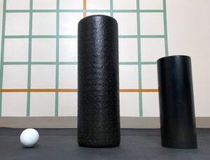
Just click on an area of the body to learn about it’s function and how to release it on your own! Before you get started, you’re going to need some basic equipment. All you need is a foam roller and a tennis ball. If that doesn’t feel like it’s massaging deep enough, you’ll need a 6″ PVC pipe and a lacrosse ball. Learn more about these tools and why they are preferred for the Deep Stretch Method by clicking the button below:
SMR Map
Select a muscle to learn about it's function and how to release it on your own!
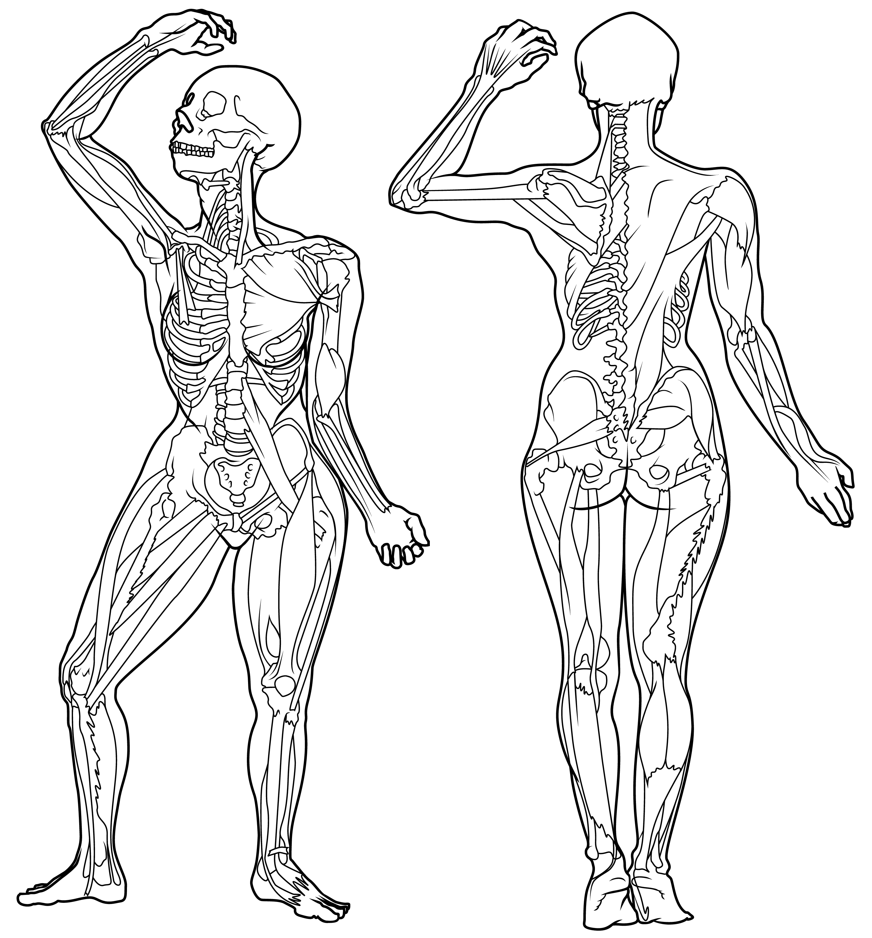
The SMR Map is a unilateral muscle anatomy chart with both anterior and posterior views. It's illustrated to have just one of each major kinesthetic driving muscle so that it's easier to understand where a muscle attaches and what it's functions are.
Click the area you want to release, and a short description of the muscle along with a link to the release video will appear in this window.
Make sure to share this resource with your friends! Thank you for visiting!
Biceps Muscles
- The biceps muscles are the two muscle heads that make up the bulk of the appearance of upper arms. Altogether, the two muscle heads along with the adjacent brachialis are commonly referred to as the 'biceps brachialis' muscle group.
- It's insertion is the radial tuberosity and it's origins are the coracoid process and superglenoid tubricle of the scapula.
- This muscle's primary action is flexion of the elbow. When this muscle contracts, it brings the forearm closer to the upper arm.
- Releasing this muscle helps improve:
- Elbow pain
- Elbow soreness
- Arm pain
- Wrist pain
- Elbow mobility
Forearm Flexors
- The forearm flexors are located in the underside of the forearm.
- They attach at their origins around the distal epicondyle of the humerus, and the insertions around the hand and digit bones.
- Their primary action is flexion of the wrist and fingers.
- Releasing this muscle helps improve:
- Carpel tunnel symptoms
- Arthritis
- Joint pain
- Elbow pain
- Local tension
Sterno-cleido-mastoid
- The Sternocleidomastoid is the most accessible muscle in the front of the neck.
- The attachments are, the origin the sternum and clavicle, and the insertion the mastoid process.
- This muscle's primary action is movement and stabilization of the head and neck.
- Releasing this muscle helps improve:
- Headaches
- Cervical alignment
- Neck pain
- Range of motion
- Flexibility
The Scalene Muscles
- The scalene muscles are deep in the front/side of the neck and pass under the collar bone (clavicle).
- The attachments are the cervical vertebrae, and first and second rib.
- This muscle group's primary action is elevation of the first and second rib during inhalation, for example. However these muscles also assist in rotation and flexion of the neck.
- Releasing this muscle helps improve:
- Headaches
- Neck pain
- Nerve pain
- Range of motion
- Fullness of breath
Pectoralis Major
- The pectoralis major is more commonly known as the 'pecs' and make up the bulk of what consider the shape of the chest.
- The attachments are, the origin the sternum and clavicle, and the insertion the proximal aspect of the humerus.
- This muscle group's primary action is to adduct and internally rotate the humerus.
- Releasing this muscle helps improve:
- Shoulder pain
- Thoracic posture
- Chest pain
- Range of motion
- Flexibility
TFL and IT Band
- The tensor fasciae latae is better known as the TFL. and the iliotibial tract is better known as the IT band. The IT band is the insertion for the TFL. The TFL is a muscle while the TFL is just connective tissue. Note: *connective tissue is more sensitive to pressure. Avoid the IT band and focus on TFL instead*
- The attachments are, the origin the iliac crest and anterior superior iliac spine (ASIS), and the insertion the IT band. The IT band then goes on to connect at the condyle of the tibia.
- This muscle group's primary action is stabilization of the hip and leg. It also assists with internal rotation and abduction of the hip.
- Releasing this muscle helps improve:
- Lower back pain
- Postural alignment
- Leg fatigue
- Range of motion
- Hip pain
Psoas
- Psoas is the one of the deepest muscles in the body. It's a deep hip flexor that brings the knee towards the chest.
- The attachments are, the lumbar spine, and the head of the femur.
- This muscle group's primary action is flexion of the leg, but it also shares a close proximity to the diaphragm. With each breath the psoas muscle and diaphragm work in synchronicity to provide stability to the front of the spine.
- Releasing this muscle helps improve:
- Lower back pain
- Postural alignment
- Hip pain
- Fullness of breath
- Flexibility
Sartorius

- Sartorius is the longest muscle in the human body. Makes sense, because it spans the entire length of the longest bone in the body, the femur.
- The attachments are, their origin the anterior superior iliac spine (ASIS), and the insertion along the proximal end of the tibia.
- This muscle group's primary action is flexion of the leg, but it also assists in external rotation of the femur. When this muscle contracts, it brings the knee closer to the body. Sitting in a chair, for example, brings the muscle into contraction.
- Releasing this muscle helps improve:
- Lower back pain
- Postural alignment
- Hip pain
- Range of motion
- Flexibility
Adductors Muscle Group
- The inner thing muscles altogether make up the adductor muscle group.
- Their attachments are; their origins along the bottom of the pelvis, and their insertions along the femur and the head of the tibia.
- This muscle group's primary action is adduction of the leg. When this muscle contracts, it brings the legs closer toward each other.
- Releasing this muscle helps improve:
- Lower back pain
- Hip mobility
- Knee pain
- Hip pain
- Flexibility
Tibialis Anterior
- The tibialis anterior is what most people would think of as the shin muscle. It's generally the muscle most effected by shin splints.
- The attachments are, the origin the upper lateral shaft of the tibia, and the insertion at the medial cuneiform and the first metatarsal.
- This muscle group's primary action is extension and inversion of the ankle.
- Releasing this muscle helps improve:
- Lower back pain
- Postural alignment
- Hip pain
- Range of motion
- Flexibility
Soleus
Extensor Hallucis
- The quadriceps muscles represent the rectus femoris, vastus medialis, lateralis, and intermedius. They are commonly referred to as the hip flexors because they are largely responsible for hip flexion.
- The attachments are, their origin wrapping around the femur and beginning at the ilium, and the insertion ends at the connective tissue surrounding the patella (knee cap).
- This muscle group's primary action is flexion of the leg. When this muscle contracts, it brings the knee closer to the body. Sitting in a chair, for example, brings the muscle into contraction.
- Releasing this muscle helps improve:
- Lower back pain
- Postural alignment
- Hip pain
- Range of motion
- Flexibility
Peroneal Muscles
- The peroneal muscles are lower leg muscles that stabilize and pronate the ankle.
- It attaches at it's origin, the proximal aspect of the fibula, and it's insertion, medial cuneiform and first metatarsal.
- This muscle's actions are pronation or eversion of the ankle, and it also assists in plantar flexion of the ankle.
- Releasing this muscle helps improve:
- Lower leg pain
- Calf cramping
- Foot/ankle posture
- Foot circulation
- Ankle mobility
Adductor Magnus
- Adductor magnus is the largest of the adductor muscle group. The muscles altogether make up the inner thigh muscles.
- Their attachments are; their origins along the bottom of the pelvis, and their insertions along the femur and the head of the tibia.
- This muscle group's primary action is adduction of the leg. When this muscle contracts, it brings the legs closer toward each other.
- Releasing this muscle helps improve:
- Lower back pain
- Hip mobility
- Knee pain
- Hip pain
- Flexibility
Rectus Femoris
- The quadriceps muscles represent the rectus femoris, vastus medialis, lateralis, and intermedius. They are commonly referred to as the hip flexors because they are largely responsible for hip flexion.
- The attachments are, their origin wrapping around the femur and beginning at the ilium, and the insertion ends at the connective tissue surrounding the patella (knee cap).
- This muscle group's primary action is flexion of the leg. When this muscle contracts, it brings the knee closer to the body. Sitting in a chair, for example, brings the muscle into contraction.
- Releasing this muscle helps improve:
- Lower back pain
- Postural alignment
- Hip pain
- Range of motion
- Flexibility
Vastus Lateralis
- The quadriceps muscles represent the rectus femoris, vastus medialis, lateralis, and intermedius. They are commonly referred to as the hip flexors because they are largely responsible for hip flexion.
- The attachments are, their origin wrapping around the femur and beginning at the ilium, and the insertion ends at the connective tissue surrounding the patella (knee cap).
- This muscle group's primary action is flexion of the hip and extension of the knee. When this muscle contracts, it brings the knee closer to the body. Sitting in a chair, for example, brings the muscle into contraction.
- Releasing this muscle helps improve:
- Lower back pain
- Postural alignment
- Hip pain
- Range of motion
- Flexibility
Triceps and Deltoid Muscles
- The deltoid and triceps muscles are two different muscle groups, but they are included in the same category because the stretch/release is the same.
- The deltoid attaches at the
- This muscle's primary action is stabilization of the hip and external rotation of the hip. When it contracts, it rotates the legs away from each other, often resulting in a 'v' shape with your feet (heels toughing). An overly dysfunctional piriformis can mimic extreme pains felt in sciatica and is referred to as piriformis syndrome.
- Releasing this muscle helps improve:
- Back pain
- Hip pressure
- Lumbar posture
- Hip posture
- Hip mobility
Quadratus Laborum
- The quadratus laborum is the deepest back muscle and is located between the hips and ribs.
- It's attachments are the origin, the crest of the pelvis, and the insertion, the L-1 through L-5 lumbar vertebrae and the 12th rib.
- This muscle's primary action is stabilization of the hips and pelvis, but it also assists with lateral flexion of the spine. When this muscle contracts, it brings one side of the hip closer to the ribcage.
- Releasing this muscle helps improve:
- Back pain
- Hip soreness
- Lower back pain
- Back/hip posture
- Lumbar mobility
Brachialis
- The brachialis is deep to the more commonly known biceps muscles. Altogether, the three muscle heads are commonly referred to as the 'biceps brachialis' muscle group.
- It's attachments are the humerus and the condyle of the ulna.
- This muscle's primary action is flexion of the elbow. When this muscle contracts, it brings the forearm closer to the upper arm.
- Releasing this muscle helps improve:
- Elbow pain
- Elbow soreness
- Arm pain
- Wrist pain
- Elbow mobility
Levator Scapulae
- The levator scapulae is one of the most commonly massaged muscle in the body. It lies beneath the trapezius muscle in the upper neck/shoulder area.
- The attachments are, the origin, the transverse processes from C-1 to C-4. And the insertion the upper medial border of the scapula.
- This muscle's primary action is to raise the medial border of the scapula, but it also assists with lateral flexion and rotation of the neck.
- Releasing this muscle helps improve:
- Headaches
- Cervical alignment
- Neck pain
- Neck and shoulder range of motion
- Shoulder pain
Rhomboid Major/Minor
- The rhomboid muscles are between the spine and shoulder blade.
- Their attachments are the insertion, the border of the scapula, and the origin, the T-2 to T-5 vertebrae.
- This muscle's primary action is to slide the shoulder blade towards the spine, but it also assists in rotation of the thoracic vertebrae.
- Releasing this muscle helps improve:
- Shoulder pain
- Shoulder stability
- Back pain
- Neck pain
- Shoulder mobility
The SITS Muscles
- The SITS muscles represent four muscles that ride the surface of the scapula.
- It's attachments are the surface of the scapula and the proximal end (base) of the humerus.
- This muscle's primary action is stabilization of the shoulder and assistance in movement of the upper arm. Releasing these muscle can be helpful for pinpoint chronic pains in the chest/neck/shoulder area.
- Releasing this muscle helps improve:
- Shoulder pain
- Shoulder stability
- Chest pain
- Neck pain
- Shoulder mobility
Forearm Extensors
- The forearm flexors are located in the topside of the forearm.
- They attach at their origins around the distal epicondyle of the humerus, and the insertions around the hand and digit bones.
- Their primary action is extension of the wrist and fingers.
- Releasing this muscle helps improve:
- Carpel tunnel symptoms
- Arthritis
- Joint pain
- Elbow pain
- Local tension
Serratus
- The serratus are the "serrated" muscled that blanket the ribcage.
- The attachments are, the origin, ribs 1 through 10. And the insertion, the anterior surface of the scapula.
- This muscle's primary action is to assist in expanding the ribcage for inhalation, but it also effects rotation and stabilization of the scapula and draws the scapula anterolaterally.
- Releasing this muscle helps improve:
- Fullness of breath
- Shoulder pain
- Back pain
- Thoracic range of motion
- Thoracic mobility
Trapezius
- The trapezius is the most commonly massaged muscle in the body. It spans the entire upper back and shoulder area.
- The attachments are, the origin the superior nuchal line, ligament nuchae, and the spinous processes and supraspinous ligaments from the occipital ridge through T-12. And the insertion the upper fibers to lateral third of posterior border of clavicle; lower to medial acromion and superior lip of spine of scapula to deltoid tubercle
- This muscle's primary action is to laterally rotate, elevate, and retract the scapula, and to also laterally flex the neck.
- Releasing this muscle helps improve:
- Headaches
- Cervical alignment
- Neck pain
- Neck and shoulder range of motion
- Thoracic flexibility
Latissimus Dorsi
- The latissimus dorsi has the widest span of any muscle in the body. It also, by far, the largest arm/shoulder/back muscle.
- It attaches at it's origin; T-7 through T-12, posterior crest of the ilium, and ribs 9 through 12. And the insertion, the proximal humerus in between the pectoralis minor and teres minor muscles.
- This muscle's primary action is internal rotation of the humerus and adduction of the arm. In sport, the lats should generally be the hardest working shoulder muscle. If you're experiencing arm soreness, it's likely because the lats aren't working hard enough.
- Releasing this muscle helps improve:
- Back pain
- Shoulder pain
- Shoulder/back posture
- Shoulder flexibility
- Shoulder mobility
Triceps and Deltoid Muscles
- The piriformis is located deep to the most likely thickest muscle in the body, gluteus maximus.
- It attaches at it's origin, the sacrum, and it's insertion, the head of the femur.
- This muscle's primary action is stabilization of the hip and external rotation of the hip. When it contracts, it rotates the legs away from each other, often resulting in a 'v' shape with your feet (heels toughing). An overly dysfunctional piriformis can mimic extreme pains felt in sciatica and is referred to as piriformis syndrome.
- Releasing this muscle helps improve:
- Back pain
- Hip pressure
- Lumbar posture
- Hip posture
- Hip mobility
Piriformis
- The piriformis is located deep to the most likely thickest muscle in the body, gluteus maximus.
- It attaches at it's origin, the sacrum, and it's insertion, the head of the femur.
- This muscle's primary action is stabilization of the hip and external rotation of the hip. When it contracts, it rotates the legs away from each other, often resulting in a 'v' shape with your feet (heels toughing). An overly dysfunctional piriformis can mimic extreme pains felt in sciatica and is referred to as piriformis syndrome.
- Releasing this muscle helps improve:
- Back pain
- Hip pressure
- Lumbar posture
- Hip posture
- Hip mobility
Gluteus Medius
- The gluteus medius is a small but hard working muscle located directly above the better known gluteus maximus.
- It attaches at it's origin, the surface of the illium, and it's insertion, the greater trochanter of the femur.
- This muscle is active during every action of the hip other than lateral flexion.
- Releasing this muscle helps improve:
- Back pain
- Hip pressure
- Lumbar posture
- Hip posture
- Hip mobility
Flexor Pollicis Longus
- The pollicis muscles are the muscles that primarily control movement in the thumb.
- The attachments for flexor pollicis longus are, their origin, the anterior surface of the radius. And the insertion, the bas of the distal phalanx of the thumb.
- This muscle group's primary action is flexion of the phalanx of the thumb. When this muscle contracts, it brings the thumb away from the palm and brings it towards the upper forearm.
- Releasing this muscle helps improve:
- Wrist pain
- Wrist mobility
- Thumb/hand pain
- Range of motion
- Flexibility
Hamstrings Muscle Group
- The hamstring muscles represent four different muscle heads. Together they make up the muscles that make up the back of your leg.
- The attachments are, their origin wrapping around the femur and beginning at the ilium, and the insertion ends at the connective tissue surrounding the patella (knee cap).
- This muscle group's primary action is extension of the hip and flexion of the knee. When this muscle contracts, it brings the knee and foot closer to the back of the body. Sitting in a chair, for example, brings the muscle into contraction.
- Releasing this muscle helps improve:
- Lower back pain
- Postural alignment
- Hip pain
- Range of motion
- Flexibility
Vastus Medialis
- The quadriceps muscles represent the rectus femoris, vastus medialis, lateralis, and intermedius. They are commonly referred to as the hip flexors because they are largely responsible for hip flexion.
- The attachments are, their origin wrapping around the femur and beginning at the ilium, and the insertion ends at the connective tissue surrounding the patella (knee cap).
- This muscle group's primary action is flexion of the hip and extension of the knee. When this muscle contracts, it brings the knee closer to the body. Sitting in a chair, for example, brings the muscle into contraction.
- Releasing this muscle helps improve:
- Lower back pain
- Postural alignment
- Hip pain
- Range of motion
- Flexibility
Hamstrings Muscle Group
- The hamstring muscles represent four different muscle heads. Together they make up the muscles that make up the back of your leg.
- The attachments are, their origin wrapping around the femur and beginning at the ilium, and the insertion ends at the connective tissue surrounding the patella (knee cap).
- This muscle group's primary action is extension of the hip and flexion of the knee. When this muscle contracts, it brings the knee and foot closer to the back of the body. Sitting in a chair, for example, brings the muscle into contraction.
- Releasing this muscle helps improve:
- Lower back pain
- Postural alignment
- Hip pain
- Range of motion
- Flexibility
Vastus Lateralus
- The quadriceps muscles represent the rectus femoris, vastus medialis, lateralis, and intermedius. They are commonly referred to as the hip flexors because they are largely responsible for hip flexion.
- The attachments are, their origin wrapping around the femur and beginning at the ilium, and the insertion ends at the connective tissue surrounding the patella (knee cap).
- This muscle group's primary action is flexion of the hip and extension of the knee. When this muscle contracts, it brings the knee closer to the body. Sitting in a chair, for example, brings the muscle into contraction.
- Releasing this muscle helps improve:
- Lower back pain
- Postural alignment
- Hip pain
- Range of motion
- Flexibility
Gastrocnemius and Achilles
- The gastrocnemius is more commonly known as the bulk of the calf muscle.
- It attaches at it's origin, the condyles of the femur, and it's insertion, the calcaneus or the heel.
- This muscle's primary action is dorsiflexion. When it contracts, the foot presses down like you would agas or break pedal.
- Releasing this muscle helps improve:
- Calf cramps
- Ankle stability
- Knee pain
- Plantar fasciitis
- Stamina
Tibialis Posterior
- The tibialis posterior is a deep muscle in the lower leg.
- The attachments are, the origin the posterior surface of tibia, posterior surface of fibula and interosseous membrane. And the insertions; the navicular tuberosity, cuneiforms, cuboid, 2-4 metatarsals, and sustentaculum tali of calcaneus.
- This muscle group's primary action is to plantarflex the foot or press the foot downward like pressing down on a gas pedal.
- Releasing this muscle helps improve:
- Ankle pain
- Ankle flexibility
- Knee pain
- Range of motion
- Flexibility
Forearm Extensors
- The forearm flexors are located in the topside of the forearm.
- They attach at their origins around the distal epicondyle of the humerus, and the insertions around the hand and digit bones.
- Their primary action is extension of the wrist and fingers.
- Releasing this muscle helps improve:
- Carpel tunnel symptoms
- Arthritis
- Joint pain
- Elbow pain
- Local tension
Occipital Ridge
- The occipital ridge is the back of the base of the skull.
- These muscles attach at their origin, the first and second vertebrae, and their insertions, the occipital ridge of the skull.
- This muscle's primary action is stabilization of the head and neck.
- Releasing this muscle helps improve:
- Headaches
- Stress
- Upper body posture
- Neck mobility
- Neck/back pain
Forearm Flexors
- The forearm flexors are located in the underside of the forearm.
- They attach at their origins around the distal epicondyle of the humerus, and the insertions around the hand and digit bones.
- Their primary action is flexion of the wrist and fingers.
- Releasing this muscle helps improve:
- Carpel tunnel symptoms
- Arthritis
- Joint pain
- Elbow pain
- Local tension
Pectoralis Minor
- The pectoralis minor is located deep to the muscle that makes up the shape of the chest, the pec major.
- It attaches at it's origin, the third fourth and fifth ribs, and it's insertion, the coracoid process of the scapula.
- This muscle's primary action is stabilization of the shoulder and assistance in inhalation. When it contracts, it helps to expand the ribcage to enable a fuller breath.
- Releasing this muscle helps improve:
- Fullness of breath
- Shoulder stability
- Upper body posture
- Headaches
- Thoracic mobility
Subscapularis
- The subscapularis is the muscle that travels from the interior surface of the scapula to the head of the humerus. It acts like a 'deeper' pec major.
- The attachments are, the origin the subscapular fossa of the scapula, and the insertion the lesser tubercle of the humerus.
- This muscle group's primary action is to adduct and internally rotate the humerus.
- Releasing this muscle helps improve:
- Shoulder pain
- Thoracic posture
- Chest pain
- Range of motion
- Flexibility
Quadratus Femoris
- Though small, the quadratus femoris is not insignificant.
- It attaches at it's origin, the ischial tuberosity, and it's insertion, the crest of the femur.
- This muscle's primary action is stabilization of the hip and external rotation of the hip. When it contracts, it rotates the legs away from each other, often resulting in a 'v' shape with your feet (heels toughing).
- Releasing this muscle helps improve:
- Back pain
- Hip pressure
- Lumbar posture
- Hip posture
- Hip mobility
Videos